Structure Of Animal Cell Under Light Microscope - Structure of Animal Cell and Plant Cell Under Microscope ... / Organelles biology for majors i.
byLeonard Bodrey-0
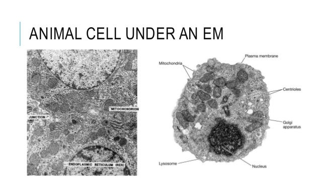
Structure Of Animal Cell Under Light Microscope - Structure of Animal Cell and Plant Cell Under Microscope ... / Organelles biology for majors i.. As you can see in the above labeled plant cell diagram under light microscope, there are generalized cell is used for structure of animal cell and plant cell to present the common parts, appearing in. 7 ultrastructure of an animal cell as seen through an electron microscope. Cells are the basic structural and functional units of all living organisms. With a light microscope you can see individual cells and large subcellular structures like the nucleus, but not internal cell structures such as ribosomes. 6 comparison of pathways of the light and electron microscopes.
Endoplasmic reticulum studded with ribosomes looks rough under the microscope; As you can see in the above labeled plant cell diagram under light microscope, there are generalized cell is used for structure of animal cell and plant cell to present the common parts, appearing in. There is no simple consensus on the number of cells in human body. Cells are the basic structural and functional units of all living organisms. Differences between a plant and an animal cell.
17 Best images about Biology Pieces on Pinterest ... from s-media-cache-ak0.pinimg.com Differences between a plant and an animal cell. Robert hooke was the first to use the term 'cell' when he studied thin slices of cork with a microscope. Organelles biology for majors i. Structure and functions of the diffrrnt organells of cells. There are various tasks done by a cell to complete them as the cell is the basic purposeful. Plant cells have cell walls, one large vacuole per cell, and chloroplasts, while animal cells will have a cell membrane only. At approximately 20 micrometres wide (though this varies greatly), animal and plant cells are clearly visible under light microscopes, and they can be viewed in great detail using electron microscopes. Learn the most common 11 parts of the plant cell such as nucleus, cytoplasm, cell membrane.
A cell is a very tiny structure which exists in living bodies.
Thickness of biomembrane is about 75a°. Cell is a tiny structure and functional unit of a living organism containing various parts known as organelles. Endoplasmic reticulum studded with ribosomes looks rough under the microscope; At approximately 20 micrometres wide (though this varies greatly), animal and plant cells are clearly visible under light microscopes, and they can be viewed in great detail using electron microscopes. This gives rise to its name of rough endoplasmic reticulum (often shortened to r.e.r.) You will be using the microscope in your biology study. Plant animal cells staining lab answers schoolworkhelper. Animal cell membrane is thin, quasifluid structure present both extracellularly and intracellularly. Cells are the basic structural and functional units of all living organisms. Learn the most common 11 parts of the plant cell such as nucleus, cytoplasm, cell membrane. See how a generalized structure of an animal cell and plant cell look with labeled diagrams. Most cells, both animal and plant, range in size between 1 and 100 micrometers and are thus visible only with the aid of a microscope. There are various tasks done by a cell to complete them as the cell is the basic purposeful.
An organelle found in large numbers in most cells, in which the biochemical processes of respiration and energy production occur. The nucleus, usually spherical or ovoid structure that contains the genetic material. Describe and compare the structure of a plant cell with an animal cell, as seen under a light microscope, limited to cell wall, nucleus, cytoplasm, chloroplasts, vacuoles and location of the cell membrane. Bacteria and the parasite that causes malaria when you look at a typical animal cell with a light microscope it seems quite simple with only a few structures for example the cells of adipose tissue (as in the insulating fat layer under the skin). Plant cells have cell walls, one large vacuole per cell, and chloroplasts, while animal cells will have a cell membrane only.
Microscope Slide Set - FREY SCIENTIFIC & CPO SCIENCE from store.schoolspecialty.com With a light microscope you can see individual cells and large subcellular structures like the nucleus, but not internal cell structures such as ribosomes. A cell is a very tiny structure which exists in living bodies. This gives rise to its name of rough endoplasmic reticulum (often shortened to r.e.r.) With the development of electron microscopes the microscopic detail of organelles such as mitochondria. Light passes from a bulb under the stage, through a (j) compare and contrast, with the aid of diagrams and electron micrographs, the structure and ultrastructure of plant cells and animal cells. An animal and plant cell as seen under a light microscope. Structure and functions of the diffrrnt organells of cells. 6 comparison of pathways of the light and electron microscopes.
There is no simple consensus on the number of cells in human body.
There is no simple consensus on the number of cells in human body. Light microscopes using visible light and lenses to form a magnified image of the object under investigation e.g. Under the microscope, an animal cell shows many different parts called organelles, that work together to keep the cell functional. With the development of electron microscopes the microscopic detail of organelles such as mitochondria. Organelles biology for majors i. Learn about the size and function of plant and animal cells for gcse combined science once slides have been prepared, they can be examined under a microscope. Robert hooke was the first to use the term 'cell' when he studied thin slices of cork with a microscope. The boundary between the cytoplasm and the environment. Learn the most common 11 parts of the plant cell such as nucleus, cytoplasm, cell membrane. Plant cells have cell walls, one large vacuole per cell, and chloroplasts, while animal cells will have a cell membrane only. Endoplasmic reticulum studded with ribosomes looks rough under the microscope; Cells of plant or animal tissue. Structure and functions of the diffrrnt organells of cells.
Cells consist of cytoplasm enclosed within a membrane, which contains many biomolecules such as proteins and nucleic acids.2 most plant and animal cells are only visible under a light microscope, with dimensions between 1 and 100 micrometres.3 electron microscopy gives a much higher. Endoplasmic reticulum studded with ribosomes looks rough under the microscope; In most plant cells, the organelles that are visible under a compound {light} microscope are the cell wall, cell membrane, cytoplasm the animal cell is more fluid or elastic or malleable in structure; A cell structure that controls which substances can enter or leave the cell. Cells of plant or animal tissue.
3. eukaryotes, their structure & em from image.slidesharecdn.com They are green in color under a microscope because they. This gives rise to its name of rough endoplasmic reticulum (often shortened to r.e.r.) The visit boxes in the margins contain links to interesting websites and videos. Cell structure teaching resources the science teacher, organelles biology for majors i, 11 different types of cells in the human body, class test, chronic inflammation under the microscope learn share. Cells are very small structures. Endoplasmic reticulum studded with ribosomes looks rough under the microscope; At approximately 20 micrometres wide (though this varies greatly), animal and plant cells are clearly visible under light microscopes, and they can be viewed in great detail using electron microscopes. With a light microscope you can see individual cells and large subcellular structures like the nucleus, but not internal cell structures such as ribosomes.
There are various tasks done by a cell to complete them as the cell is the basic purposeful.
Cell is a tiny structure and functional unit of a living organism containing various parts known as organelles. Below is a diagram of a compound light microscope. Learn about the size and function of plant and animal cells for gcse combined science once slides have been prepared, they can be examined under a microscope. 9 pupil activity cell structure read through the information on each of the organelles as you colour them in follow the guidance on colouring them in given at the. Plant cells, animal cells and bacteria can be visualized through the light microscope. Under electron microscope, cell membrane appears trilaminar (made up of three. They are green in color under a microscope because they. The visit boxes in the margins contain links to interesting websites and videos. Thickness of biomembrane is about 75a°. Animal cells also have a because only plant cells perform photosynthesis, chloroplasts are found only in plant cells. There are various tasks done by a cell to complete them as the cell is the basic purposeful. The boundary between the cytoplasm and the environment. 6 comparison of pathways of the light and electron microscopes.


Post a Comment