Plant Cell Diagram Under Light Microscope : What Are The Visible Plant Animal Cell Organs On Light Microscope Quora - Juicy green plant cells under the microscope.
byLeonard Bodrey-0
Plant Cell Diagram Under Light Microscope : What Are The Visible Plant Animal Cell Organs On Light Microscope Quora - Juicy green plant cells under the microscope.. Here's a diagram of a plant cell: In truth, there are still features of plant and animal appearance —under a microscope, normal cells and cancer cells may look quite different. Microscopy and the interpretation of cell structures. Structure of animal cell and plant cell under microscope + diagrams. The diagram is very clear, and labeled;
Turn the coarse focus so that the stage is as close to the objective lens as possible. Plant cell surface of leaf under light microscope. Labeled diagram of plant cell, created with biorender.com. A micrograph is a photo or digital image taken through a microscope to show a magnified image of a specimen. Plant cells are eukaryotic cells present in green plants, photosynthetic eukaryotes of the kingdom plantae.
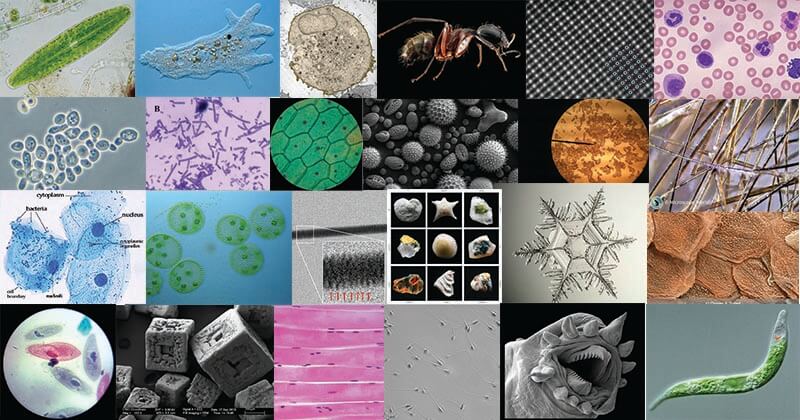
How These 26 Things Look Like Under The Microscope With Diagrams from microbenotes.com Chlorophyll, which gives plants their green color, enables them to use sunlight to convert water and carbon. Dreamstime is the world`s largest stock photography community. Cell is a tiny structure and functional unit of a living organism containing various parts known as organelles. Typical animal cell (membranes) [light m… Use them in commercial designs under lifetime, perpetual & worldwide rights. Structure of animal cell and plant cell under microscope + diagrams. Light photomicrograph of helianthus stem cross section seen through microscope. Plant cell surface of leaf under light microscope.
Mount a complete leaf of elodea in water on a slide and examine under high power of the microscope.
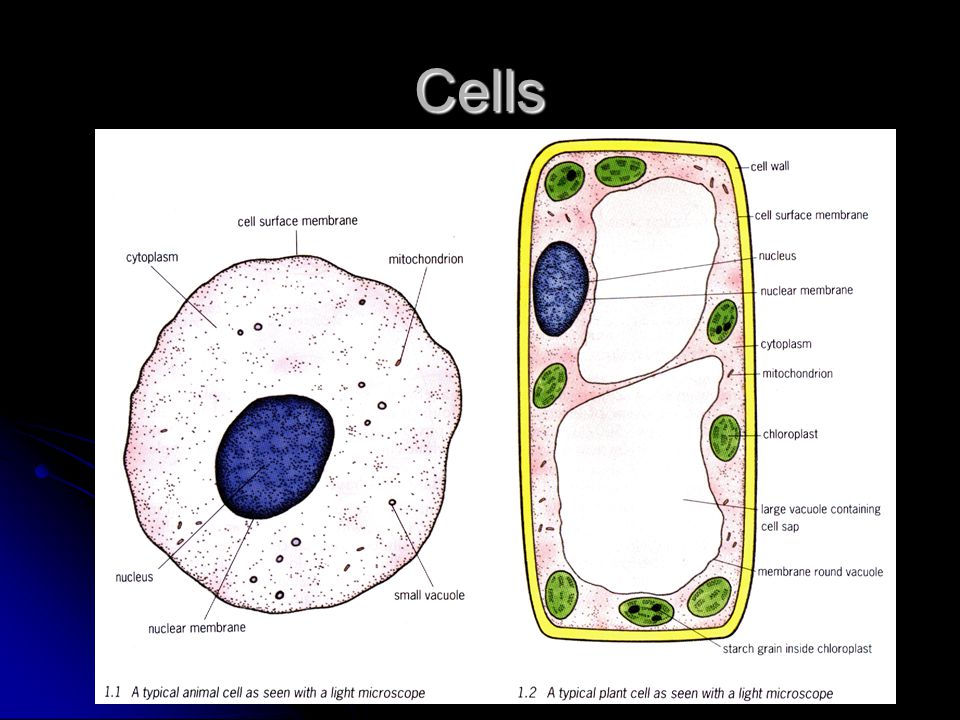
However, plant cells have a rigid experiment 1. Vector low poly solar power plant and city. Here's a photo of a plant cell under an electron microscope. Dreamstime is the world`s largest stock photography community. Describe and compare the structure of a plant cell with an animal cell, as seen under a light microscope, limited to cell wall, nucleus, cytoplasm, chloroplasts, vacuoles and location of the cell membrane. Pine mature wood cross section. The condenser focuses the light through the object. Plant cells have cell walls, one large vacuole per cell, and chloroplasts, while animal cells will have a cell membrane only. Study the two diagrams of plant and animal cells below. A few cell organelles can be seen when a plant cell is viewed under a light microscope. The microscope is perhaps one of the most fundamentally important pieces of equipment that you will use in the laboratory environment. Learn the structure of animal cell and plant cell under light microscope. Purple colored, large epidermal cells of an onion oyster plant cells.
Learn the structure of animal cell and plant cell under light microscope. Plant cells are eukaryotic cells present in green plants, photosynthetic eukaryotes of the kingdom plantae. To make observations and draw scale diagrams of cells. Their distinctive features include primary cell walls containing cellulose, hemicelluloses and pectin, the presence of plastids with the capability to perform photosynthesis and store starch. The condenser focuses the light through the object.
Plant Cell Light Microscope Diagram Auto Ken from images.slideplayer.com Cells of plant or animal tissue. The diagram is very clear, and labeled; Typical animal cell (membranes) [light m… Light microscope slide with microsection of an evergreen conifer in. The diagram is very clear, and labeled; A few cell organelles can be seen when a plant cell is viewed under a light microscope. Light microscopes using visible light and lenses to form a magnified image of the object under investigation e.g. Resolving power is the ability to distinguish between separate things which are close to each other.
Purple colored, large epidermal cells of an onion oyster plant cells.
It's a thin slice with light microscopy i can simply scrape some cells from my cheek smear them on a slide and look at. Light photomicrograph of helianthus stem cross section seen through microscope. Your plant cells under microscope stock images are ready. Pine mature wood cross section. Magnification, however, is not the most important issue in microscopy. The condenser focuses the light through the object. Vector low poly solar power plant and city. Under ordinary light microscope only few cell organelles like mitochondria, golgi complex however, under electron microscope, several other cytoplasmic organelles such as endoplasmic plastids are present only in plant cells (not in animal cells). Most light microscopes will enlarge a specimen up to 1000 times (1000x) but the electron microscope enlarge the specimen 250. Describe and compare the structure of a plant cell with an animal cell, as seen under a light microscope, limited to cell wall, nucleus, cytoplasm, chloroplasts, vacuoles and location of the cell membrane. A micrograph is a photo or digital image taken through a microscope to show a magnified image of a specimen. Cell is a tiny structure and functional unit of a living organism containing various parts known as organelles. The high resolving power makes the electron microscope a very important research tool in microbiology.
As biology 9700 chapter cell structure prepared peter ting chapter cell structure unit the microscope in cell studies compare the structure of typical. Plant and animal cells are similar, consisting of a protoplast bounded by a cell membrane. Typical animal cell (membranes) [light m… Cell is a tiny structure and functional unit of a living organism containing various parts known as organelles. However, plant cells have a rigid experiment 1.
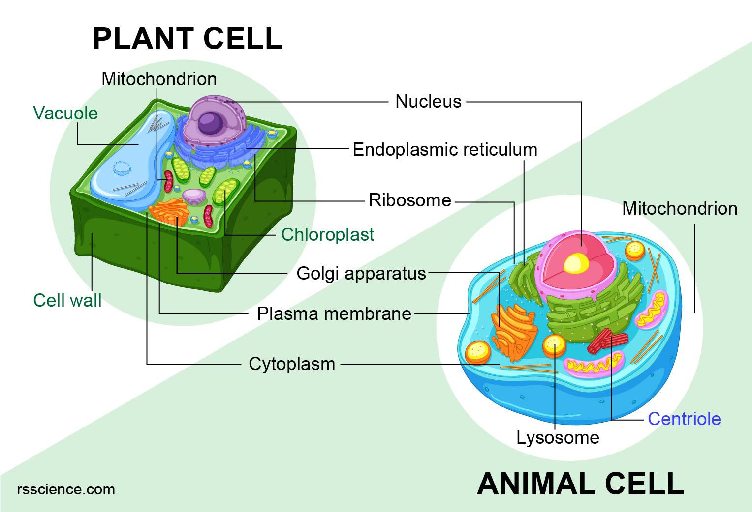
Animal Vs Plant Cells Similarities Differences Chart And Examples Rs Science from rsscience.com As you can see in the above labeled plant cell diagram under light microscope, there generalized cell is used for structure of animal cell and plant cell to present the common parts, appearing in. Cell is a tiny structure and functional unit of a living organism containing various parts known as organelles. Plant cell surface of leaf under light microscope. A micrograph is a photo or digital image taken through a microscope to show a magnified image of a specimen. Study the two diagrams of plant and animal cells below. Plant cells are the basic unit and building blocks of life in organisms of the kingdom plantae. Use them in commercial designs under lifetime, perpetual & worldwide rights. Cell is a tiny structure and functional unit of a living organism containing various parts known as organelles.
However, plant cells have a rigid experiment 1.
Mount a complete leaf of elodea in water on a slide and examine under high power of the microscope. In truth, there are still features of plant and animal appearance —under a microscope, normal cells and cancer cells may look quite different. Plant cells are the basic unit and building blocks of life in organisms of the kingdom plantae. Here's a photo of a plant cell under an electron microscope. Plant cell is an eukaryotic cell primarily involved in photosynthesis and having its genomic content some of these differences can be clearly understood when the cells are examined under an electron microscope. Transport proteins modified by the golgi body outside of the cell. Image:plant cell seen under electron microscope. You should not look through the microscope to do this. Turn the coarse focus so that the stage is as close to the objective lens as possible. Dreamstime is the world`s largest stock photography community. These include the cell wall, cell membrane, nucleus, chloroplasts. As biology 9700 chapter cell structure prepared peter ting chapter cell structure unit the microscope in cell studies compare the structure of typical. The high resolving power makes the electron microscope a very important research tool in microbiology.

Post a Comment